facilities
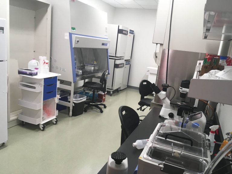
The Cellular Engineering and Mechanobiology Lab is located at the Translational Medicine Institute (TMI) at the south campus which is in closed proximity with the Flint Animal Cancer Center, Veterinary Diagnostic Laboratory, Veterinary Teaching Hospital, Orthopaedic Research Center and the Orthopaedic Bioengineering Research Laboratory. Therefore we have easy access to plenty of equipment and facilities in our vicinity. Besides, we have access to several core facilities in the main campus. Below you can find the equipment that is primarily used by our lab. The five primary dedicated spaces for our lab are: 1) Cell and tissue culture room, 2) Confocal microscopy room, 3) Molecular biology station, 4) Gel station, 5) General Lab bench area. The other facilities that we have access to in the same wet lab area and in the contiguous area (inside TMI building or adjacent buildings) are also listed in separate sections.
Cell and Tissue Culture Room
- Biosafety cabinets
- Several incubators
- A floor top centrifuge
- 4 Degree centigrade refrigerator
- -20 Degree centigrade freezer
- -80 Degree centigrade freezer (in the contiguous freezer farm)
- Cryogenic storage dewar (in the contiguous freezer farm)
- Centralized liquid nitrogen filling pipeline
- Dual water bath (two independent temperature can be set)
- Cell culture microscope with 4X-40X objectives and a color camera; computer with dedicated image acquisition software
- Dissection microscope with a color camera, computer with dedicated image acquisition software
- Countess II FL automated cell counter
- smaller items: vortex mixers, minicentrifuge, pipette sets etc.
- Access to tri-gas incubators for hypoxic cell culture in the contiguous cell and tissue culture room, separate bacterial culture room


Confocal Microscope
We recently procured a high end laser scanning confocal microscope – thanks to the generous contribution by the Translational Medicine Institute, Office of the Vice President of Research and several other sources. It is a part of the microscopy core facility and PI Dr. Soham Ghosh is the faculty in charge for this equipment. It will be installed in the microscopy room sometime mid-November. It opens a plethora of amazing capabilities which enable us to do live cell/ tissue high resolution spatiotemporal imaging, deep tissue imaging (not as good as a two photon, but better than many standard confocal), fast imaging of large samples such as tissues, environmental control for long-term live event visualization such as chromatin remodeling, protein/ mRNA movement, intracellular structure and organelle reorganization and cell migration, multiple color imaging from the same sample – thanks to the spectral imaging ability), FRET imaging etc. Therefore this microscope enables us to ask technically challenging and ambitious scientific questions. The detailed specifications are as follows.
Zeiss LSM 980 – 34 channel PMT system and TPMT with (i) Airyscan 2 with Multiplex for super resolution and fast imaging, (ii) Laser lines: 405, 488, 561 and 639 nm diode lasers ( imaging possible with many more colors simultaneously from the same sample – thanks to the spectral imaging), (iii) Axio Observer 7 inverted body with definite focus, (iv) motorized XYZ piezo for fast imaging, (v) Environmental incubation control system (completely encasing the microscope), (vi) objectives: 2.5X, 10X, 20X, 40X and 63X oil, (vii) Full system computer/ workstation and separate offline computer/ workstation.

Molecular Biology Station
- Quantstudio 3 Real Time PCR system, with dedicated computer and software
- Miniamp thermal cycler with touch screen
- Nanodrop One UV-Vis spectrophotometer with cuvette (touch screen)
- Sorvall Legend 21R temperature controlled microcentrifuge
- Neon transfection system
- Smaller items: vortex mixers, minicentrifuge, pipette sets etc


Gel Station
- iwestern workflow system (mini gel tank, iblot, ibind)
- Horinzontal gel electrophoresis tank
- Power source for gel electrophoresis

General Lab Bench Area
- Lab benches and cabinets
- Gravity oven
- Orbital shaker
- Rotator, shaker, dry bath
- Smaller items: scales, vortex mixers, minicentrifuge, pipette sets, vacuum chamber, laboratory corona handheld corona treater etc.


Access to Facility in Wet Lab Area (Common and Other PI)
- Autoclaves
- Ice making machine
- Mili-Q water stations
- Untracentrifuge
- 4 degree centigrade room, dedicated chemical room, fume hoods
- Cleanroom for nanofabrication
- Plate reader
- Miscellaneous equipment : gel doc, pH meter, untrasonic bath
- Cryotome, Immunohistochemistry facility
- Flow cytometry facility
- Fluorescence microscope (widefield)
Access to Facility in Contiguous Buildings and Inside TMI
- Spinning disk confocal microscope (at Flint Animal Cancer Center)
- Chemiluminescence gel documentation system (at Flint Animal Cancer Center)
- Mechanical characterization facility (at Orthopaedic Bioengineering Research Center)
- Histology facility (at Orthopaedic Bioengineering Research Center/ Orthopaedic Research Center)
- Small animal husbandry facility (inside TMI)
- Small and large animal imaging facility – CT, MRI (inside TMI)
- Small and large animal surgical suite (inside TMI)
Core Facilities on Main Campus That We Use
- Microscope Imaging Network for laser scanning confocal fluorescence microscopy, super resolution microscopy
- Central Instrument Facility for mass spectrometry, X-ray diffraction, Scanning Electron Microscopy, Transmission Electron Microscopy, Atomic Force Microscopy
- Flow cytometry facility
- Proteomics and metabolomics facility
- Molecular quantification core
- Next generation sequencing and genomics
- The histone source – protein purification facility
Student and Faculty Offices
Students and faculty have their own offices adjacent to the lab space. There are several conference rooms that we can use, some having outstanding view of the Rockies.

